Common anions (ClO4-, SCN- and NO3-) reduce production of TH through competitive inhibition at the Sodium Iodide Symporter (NIS). The NIS is the transport molecule that normally transfers iodine into the thyroid gland for incorporation into T4 and T3. Perchlorate is the most powerful THD with a 30x greater affinity for the NIS than does its normal target, iodine. It is about ten times as powerful as SCN- in this respect. Nitrate is a relatively weak inhibitor of iodine uptake and is believed to be 300x lower than ClO4-. However, concentrations in drinking water may be magnitudes higher. There appears to be no synergistic mechanism, and total exposure of each anion will be additive once the relative differences in strength are taken into account. All have been associated with altered TSH in infants.
The normal response to low T4/T3 is to stimulate hypothalamic release of TRH which prompts release of TSH. In a healthy individual, free of auto-immune or other thyroid disease, concentrations of TSH and T4/T3 are markers of low iodine intake, or of exposure to THDs. An individual with low iodine intake and/or high THD-exposure would be expected to have elevated TSH and/or diminished T4/T3. TSH is significantly elevated in perchlorate-exposed women and infants (BLOUNT). Any change in thyroid hormone concentrations is considered an indication of adverse effect for thyroid-hormone disrupting chemicals. Altered thyroid hormone status is, at present, the most important indicator of adverse effect in humans for thyroid-disrupting chemicals.
Glenn SM, Lester LJ. 2010. An analysis of the relationship between land use and arsenic, vanadium, nitrate and boron contamination in the Gulf Coast aquifer of Texas. Journal of Hydrology. 389: 214-226. doa: 10.1016/j.jhydrol.2010.06.002.
Tonacchera M, Pinchera A, Dimida A, Ferrarini E, Agretti P, Vitti P, Santini F, Crump K, Gibbs J. 2004. Relative potencies and additivity of perchlorate, thiocyanate, nitrate, and I- on the inhibition of radioactive I- uptake by the human sodium I- symporter. Thyroid 14(12): 1012-19.
Cao Y, Blount BC, Valentin-Blasini L, Bernbaum J, Phillips TM, Rogan WJ. 2010. Environmental Health Perspectives. 118(9): 1332-1337. DOI: 10.1289/ehp.0901736.
Miller MD, Crofton KM, Rice DC, Zoeller RT. 2009. Thyroid-Disrupting Chemicals: Interpreting Upstream Biomarkers of Adverse Outcomes. Environmental Health Perspectives. 117(7) 1033-1041,
Showing posts with label Kirk. Show all posts
Showing posts with label Kirk. Show all posts
Monday, March 7, 2011


Tuesday, March 1, 2011
Mild depression of maternal thyroid hormone adversely effects neurodevelopment of infants.
Maternal prenatal iodine deficiency hypothyroidism or hypothyroxinemia impairs intelligence and function of offspring, even when the deficiency is mild.[1],[2],[3] For neonates TSH, rather than T4 is the preferred correlate for measures of neurological/cognitive outcome. [4] Normal infants with TSH levels in the upper quartile of the normal range (4.19-17.0 mU/I)TSH score lower on tests of cognitive and executive function than those in the second quartile (2.05-2.95 mU/I). High-normal TSH has also been negatively correlated with poorer performance on measures of cognitive function and ADHD.[5] Children with urinary I levels <100 µg/L have lower IQ and higher prevalence of behavioral disorders than higher I excreting peers,while mild I deficiency in women is correlated with attention deficit hyperactivity disorders in their children.[6] An increase in congenital hypothyroidism in neonates has been noted in the US. [7],[8] Associations between transient maternal hypothyroxinemia (defined as fT4 lower than the 10th percentile) and exposure to TH-disrupting agents and autism have also been made.[9] The question of whether, and to what degree, iodine-uptake inhibitors increase rates of impairment is unanswered.
[1] Berbel P, Mestre JL, Santmira A, Palazon I, Franco A, Graells M, Gonzalez-Torga A, de Escobar GM. 2009. Delayed neurobehavioral development in children born to pregnant women with mild hypothyroxinemia during the first month of gestation: the importance of early iodine supplementation. Thyroid. 19(5): 511-519. DOI: 10.1089/thy.2008.0341
[2] Pop VJ, Kuijpens JL, van Baar AL, Verkerk G, van Son MM, de Vijlder JJ, Vulsma T, Wiersinga WM, Drexhage HA, Vader HL. 1999. Low maternal free thyroxine concentrations during early pregnancy are associated with impaired psychomotor development in infancy. Clinical Endocrinology. 50(2): 149-155.
[3] Vermiglio F, Lo Presti VP, Moleti M, Sidoti M, Tortorella G, Scaffidi G, Castagna MG, Mattina F, Violi MA, Crisa A, Aremisia A, Trimachi F. 2004. Attention deficit and hyperactivity disorders in the offspring of mothers exposed to mild-moderate iodine deficiency: A possible novel iodine deficiency disorder in developed countries. Journal of Clinical Endocrinology and Metabolism. 89(12): 6054-6060.
[4] Oken E, Braverman LE, Platek D, Mitchell ML, Lee SL, Pearce EN. 2009. Neonatal thyroxine, maternal thyroid function and child cognifition. The Journal of Clinical Endocrinology and Metabolism. 94(2): 497-503. doi:10.1210/jc.2008-0936
[5] Pederol-Ivarez M, Ribas-Fito N, Torrent M, Julvez J, Ferrer C, Sunyer J. 2007. TSH concentration within normal range is associated withy cognitive function and ADHD symptoms in healthy preschoolers. Clinical Endocrinology. 66: 890-898. (doi:10.1111/j.1365-2265.2007.02871.x)
[7] Harris KB, Pass KA. 2007. Increase in congenital hypothyroidism in New York State and in the United States. Molecular Genetics and Metabolism. 91: 268-277.
[8] Hinton CF, Harris KB, Borgfeld L, Drummond-Borg M, Eaton R, Lorey F, Therrell BL, Wallace J, Pass KA. 2010. Trends in incidence rates of congenital hypothyroidism related to select demographic factors: data from the United States, California, Massachusettes, New York and Texas. Pediatrics. 125: S37-S47.
[9] Roman G. 2007. Autism: Transient in utero hypothyroxinemia related to maternal flavonoid ingestion during pregnancy and to other environmental antithyroid agents. Journal of Neurological Sciences. 262(1-2): 15-26.
Monday, February 21, 2011
Fetal and neonatal thyroid function, review and summary of significant new findings.
Thyroid stimulating hormone (TSH) is the hormone most commonly analyzed when an infant is tested for congenital hypothyroidism (CH). Since tests for CH are required by state law, data for infant TSH is often available and no additional blood draw is required for the proposed investigation of the effects of iodine-uptake inhibitor-exposure on infant thyroid hormone (TH) levels. However, a number of factors need to be considered when correlating exposures with TSH. Neonatal TSH rises at birth and then subsides to over the next several weeks. This makes standardization of timing of sample collection important, as failure to do so creates a powerful potential confounder when comparing TSH levels among cohorts or even in determining if an individual infant’s TSH is elevated or in the process of stabilizing.
There is no national protocol for testing for congenital hypothyroidism, and the best approach is a topic of current debate and investigation.[1] Texas uses neonatal thyroxine (T4) levels to test newborns for congenital hypothyroidism because these levels are somewhat more stable, peaking at 24 hours after birth,[2] and because the assay is less expensive than that for TSH (Texas Department of Health 2011, Personal Communication). Texas State Law requires that all infants receive a T4 test at least 24 hours after birth, but before discharge from the hospital. It is also mandated that all infants receive a second test 1-2 weeks after birth. Infants whose T4 levels fall in the bottom 10th centile receive a confirmatory TSH test.[3][MSOffice1] , which would add additional data and statistical power to the proposed study.
[1] Raymond J, LaFranchi SH. 2010. Fetal and neonatal thyroid function: review and summary of significant new findings. Current Opinion in Endocrinology, Diabetes and Obesity. 17:1-7.
[2] Brown RS, Huang SA, Fisher D. 2000. The maturation of thyroid function in the perinatal period and during childhood. In: Braverman LE, Utiger RD, editors, Werner and Ingbar’s The Thyroid. Philadelphia, PA. Lippincott Williams and Wilkins. pp. 1013-1028.
[3] Texas Department of State Health Services. 2011. Newborn Screening Case Mangement. Congenital hypothyroidism: Guidelines for Professionals. http://www.dshs.state.tx.us/newborn/c_thyro.shtm
Saturday, January 22, 2011
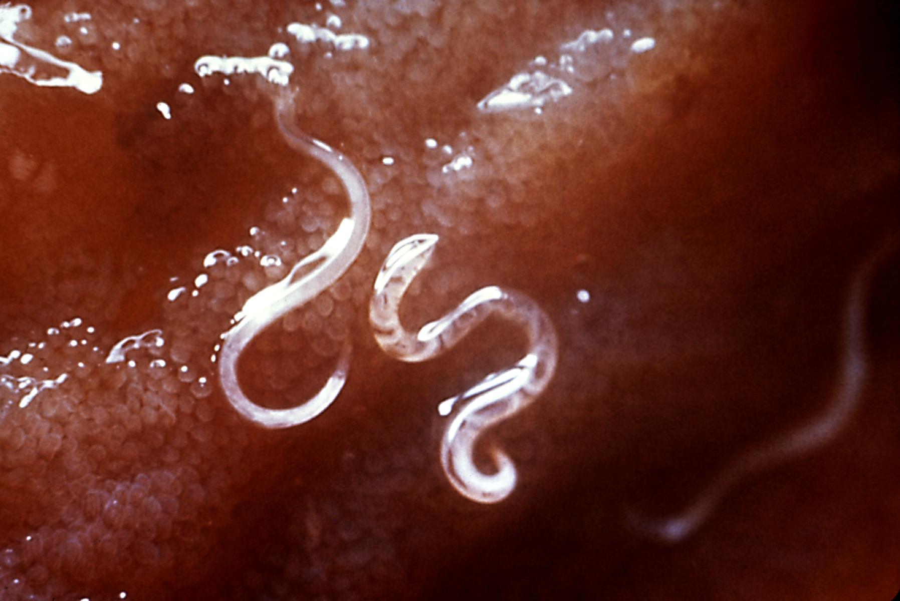
Did intestinal parasites protect us from stomach aches? Draft
Autoimmune diseases are troubling, and often frustrating and painful for health care practitioners and patients. As far as I know, celiac disease is the only autoimmune disorder with a specific and identified trigger: the protein gluten. It has also been pointed out that the incidence of Celiac Disease, the formal name for gluten-triggered autoimmune reactions in the bowel, appears to have been increasing. Some have claimed that humans are not adapted to consumption of plant proteins like gluten, and that inflammatory bowel disorders are the result of the development of agriculture. However, there are a few other possibilities. One is that autoimmune disorders in general have been increasing and that Celiac disease is not increasing at a uniquely faster rate.
A major hypothesis in the development of allergies (inappropriate immune responses to non-pathogens, such as glutten or pollen or dog hair) is lack of exposure to potential allergens early in life. This, perhaps, is the root of the problem: soap, lysol, and a generally high level of cleanliness. Also, a huge change in human habit is the widespread introduction of infant formula to replace breast milk, and early introduction of other foods into infant diets. The ideal window of exposure to gluten is now considered to be between 4 and 7 months and that exposure should be concurrent with continuing breast milk consumption. Introduction to wheat cereals during this period is thought to be protective against celiac disease. Those raising their young children on gluten-free diets in the hope of protecting them from celiac disease, or the adverse effects of inflammation in general, may be taking the wrong route. (See Pinier et al. 2010 and Lopez-Serrano et al. 2010). Let them get a little dirty
Another possiblity, proposed by Elliot and Weinstock (2009) is the current rarity of helminth infection in the Western World. These authors propose that helminth infection was protective against autoimmune diseases like celiac disease by stimulating immune circuits that lower inflammation. I suppose if one were to choose between a gluten-free diet and helminth infection, most would go with the gluten-free diet. However, before you consider loss of bread as the price some pay for the protection of all against intestinal parasites, note that Elliot and Weinstock also propose exploration of the use of helminths for clinical treatment of auto-immune disease.

Elliott, DE and Weinstock JV (2009). Inflammatory bowel disease and the hygiene hypothesis: an argument for the role of helminths Progress in Inflammation Research, 149-178 DOI: 10.1007/978-3-7643-8903-1_9
Pinier M, Fuhrmann G, Verdu E, & Leroux JC (2010). Prevention measures and exploratory pharmacological treatments of celiac disease. The American journal of gastroenterology, 105 (12) PMID: 20877349
López-Serrano P, Pérez-Calle JL, Pérez-Fernández MT, Fernández-Font JM, Boixeda de Miguel D, & Fernández-Rodríguez CM (2010). Environmental risk factors in inflammatory bowel diseases. Investigating the hygiene hypothesis: a Spanish case-control study. Scandinavian journal of gastroenterology, 45 (12), 1464-71 PMID: 20704469
A major hypothesis in the development of allergies (inappropriate immune responses to non-pathogens, such as glutten or pollen or dog hair) is lack of exposure to potential allergens early in life. This, perhaps, is the root of the problem: soap, lysol, and a generally high level of cleanliness. Also, a huge change in human habit is the widespread introduction of infant formula to replace breast milk, and early introduction of other foods into infant diets. The ideal window of exposure to gluten is now considered to be between 4 and 7 months and that exposure should be concurrent with continuing breast milk consumption. Introduction to wheat cereals during this period is thought to be protective against celiac disease. Those raising their young children on gluten-free diets in the hope of protecting them from celiac disease, or the adverse effects of inflammation in general, may be taking the wrong route. (See Pinier et al. 2010 and Lopez-Serrano et al. 2010). Let them get a little dirty
Another possiblity, proposed by Elliot and Weinstock (2009) is the current rarity of helminth infection in the Western World. These authors propose that helminth infection was protective against autoimmune diseases like celiac disease by stimulating immune circuits that lower inflammation. I suppose if one were to choose between a gluten-free diet and helminth infection, most would go with the gluten-free diet. However, before you consider loss of bread as the price some pay for the protection of all against intestinal parasites, note that Elliot and Weinstock also propose exploration of the use of helminths for clinical treatment of auto-immune disease.
Elliott, DE and Weinstock JV (2009). Inflammatory bowel disease and the hygiene hypothesis: an argument for the role of helminths Progress in Inflammation Research, 149-178 DOI: 10.1007/978-3-7643-8903-1_9
Pinier M, Fuhrmann G, Verdu E, & Leroux JC (2010). Prevention measures and exploratory pharmacological treatments of celiac disease. The American journal of gastroenterology, 105 (12) PMID: 20877349
López-Serrano P, Pérez-Calle JL, Pérez-Fernández MT, Fernández-Font JM, Boixeda de Miguel D, & Fernández-Rodríguez CM (2010). Environmental risk factors in inflammatory bowel diseases. Investigating the hygiene hypothesis: a Spanish case-control study. Scandinavian journal of gastroenterology, 45 (12), 1464-71 PMID: 20704469
Wednesday, January 19, 2011
TheseTheses
Atrazine
Congratulations to Walid D. Fakhouri, Joseph L. Nuñez, and Frances Trail for their recently published work on reduction of growth hormone production by the herbicide Atrazine. Growth hormone is important, as will sound obvious, because it plays a key role in growth (cell proliferation) but also because it influences production of many other hormones including those important in sexual development and reproduction. Changes in growth hormone produce a range of effects that extend far beyond body size.
The battles over Atrazine have been politically intense and, at times, highly entertaining to those on the sidelines. It was banned in Europe in 2004, but remained in use in the US after studies were provided to the EPA through Syngenta, the company that manufactures Atrazine, that contradicted studies by independent scientist Tyrone Hayes at the University of California at Berkeley.
Understanding Atrazine's mechanism of action will be invaluable in making informed well-reasoned decisions for protection of human health and the environment.
Fakhouri, W., Nuñez, J., & Trail, F. (2010). Atrazine Binds to the Growth Hormone–Releasing Hormone Receptor and Affects Growth Hormone Gene Expression Environmental Health Perspectives, 118 (10), 1400-1405 DOI: 10.1289/ehp.0900738
The battles over Atrazine have been politically intense and, at times, highly entertaining to those on the sidelines. It was banned in Europe in 2004, but remained in use in the US after studies were provided to the EPA through Syngenta, the company that manufactures Atrazine, that contradicted studies by independent scientist Tyrone Hayes at the University of California at Berkeley.
Understanding Atrazine's mechanism of action will be invaluable in making informed well-reasoned decisions for protection of human health and the environment.
Fakhouri, W., Nuñez, J., & Trail, F. (2010). Atrazine Binds to the Growth Hormone–Releasing Hormone Receptor and Affects Growth Hormone Gene Expression Environmental Health Perspectives, 118 (10), 1400-1405 DOI: 10.1289/ehp.0900738
Subscribe to:
Posts (Atom)